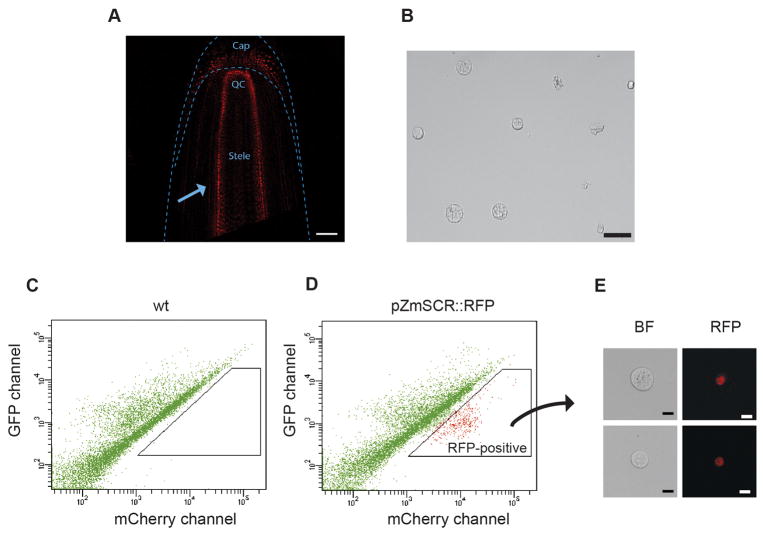
Figure 2.
Sorting of RFP-positive maize endodermal cells. Transgenic reporter lines expressing RFP in the root endodermal nuclei are screened under the microscope to ensure only RFP-expressing plants are used for protoplasts preparation. The arrow points to the layer of fluorescent endodermal cells. Scale bar, 100 μm (A). Root tips are digested, and protoplasts are inspected and counted after filtration. Scale bar 50 μm (B). A control population of protoplasted root tips (No RFP expression) is loaded into the FACS machine. A scatter plot of red versus green emission channels is generated. The resulting pattern is due to the protoplasts autofluorescence (C). Protoplasts from RFP-expressing roots show a distinct pattern when viewed on the same scatter plot. An RFP-positive population clearly appears shifted higher in the red channel axis (D). A small sample of the collected RFP-positive protoplasts are examined under the confocal microscope to ensure they have the expected fluorescence. Scale bar 10 μm (E).

