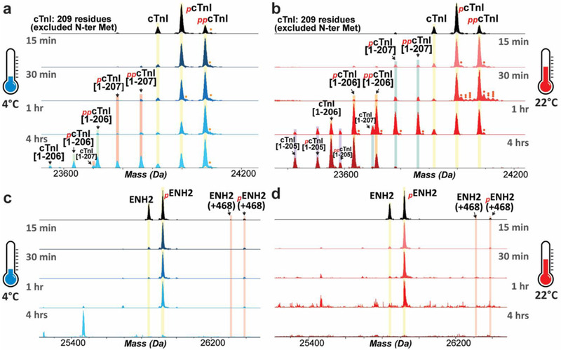
Figure 2. Rapid increase in the relative abundances of phosphorylated and degraded proteoforms of cTnI and ENH2 following increased temperature.

Top-down mass spectra show that hyper-phosphorylation and truncation of cTnI occurred after tissue incubation at 4 °C (a) or 22 °C (b). Increased phosphorylation and degradation of ENH2 upon tissue defrosting at 4 °C (c) or 22 °C (d). Single, double, and triple circles denote singly-, doubly- and triply-oxidated proteoforms, respectively.
