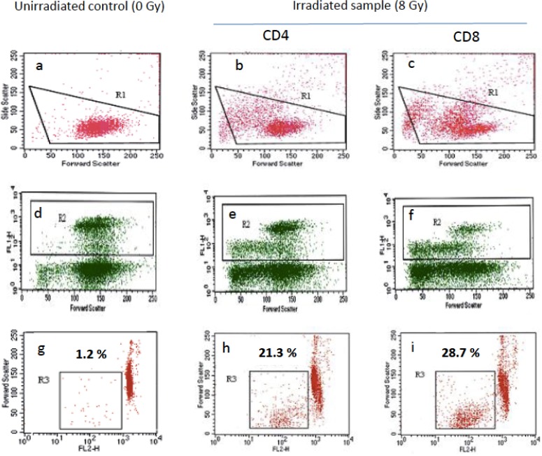
Figure 2. Radiation-induced lymphocyte apoptosis (RILA) flow cytometry profile.
Lymphocytes were selected (R1) from double-scatter dot plots (a-c) i.e. cell granularity (side scatter) and cell size (forward scatter). CD8 T-lymphocytes (R2) were then selected from R1 gated area as anti-CD4 or anti-CD8 FITC-conjugated antibody fixing cells (FL1H) (d-f). CD4 (h) or CD8 (i) T-lymphocyte apoptosis rate (R3) was then calculated from the fraction of cells from R2 gated area with reduced size (forward scatter) and reduced DNA content i.e low propidium iodide labeling (FL2H). RILA was calculated after deduction of apoptosis rate of unirradiated controls (g) from irradiated samples (h, i): respectively 20.1 and 27.5 % for CD4 and CD8 RILA in the present illustrative example.

