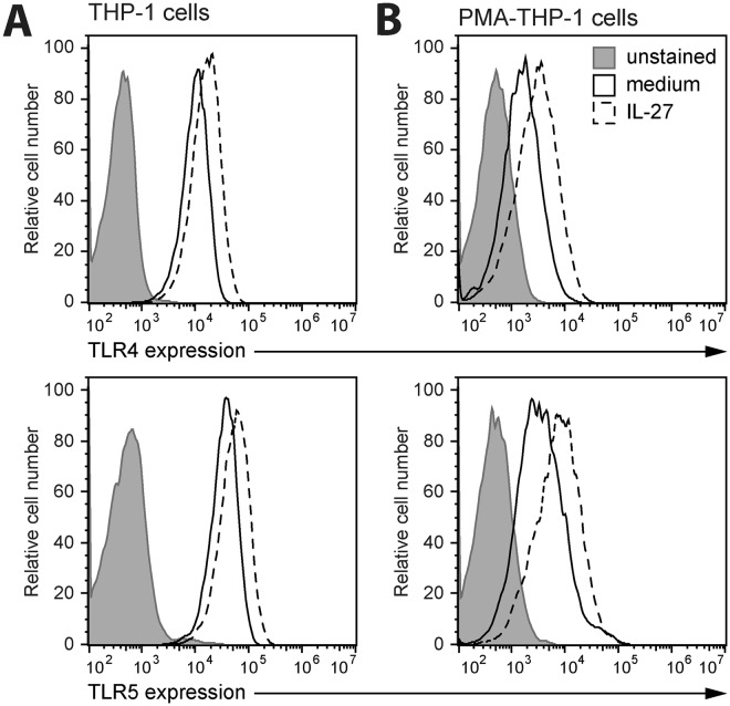
Figure 3.
Stimulation with IL-27 increased TLR4 and TLR5 expression in monocytes and macrophages. THP-1 cells (A) and PMA-THP-1 cells (B) were stimulated with or without IL-27 (50 ng/ml) for 16 hours. Cells were stained with anti-human TLR4 (top panels) or TLR5 (bottom panels) antibodies for receptor expression quantification by flow cytometry. Unstained cells were acquired to quantify autofluorescence of each cell type. Data are representative of at least three independent replicate experiments.

