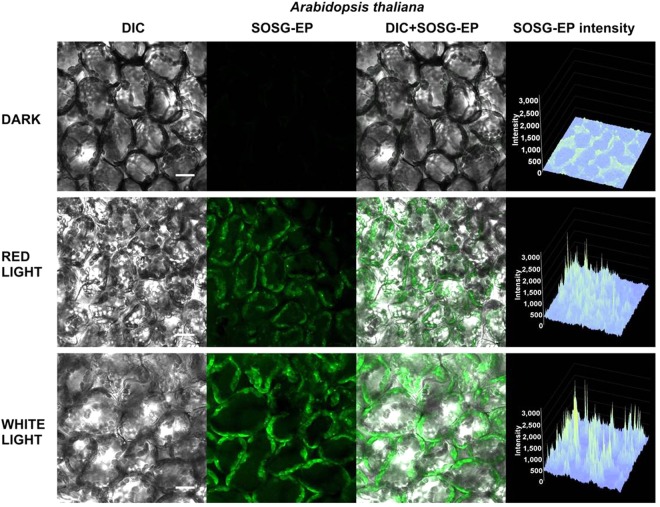
Figure 11.
Effect of red and white light on singlet oxygen imaging in Arabidopsis leaves. Arabidopsis leaves were treated in 50 μM SOSG for 30 min either in dark or exposed to red/white light. For each treatment following images are presented (from left to right): Nomarski DIC, SOSG-EP fluorescence (λem = 505–525 nm), combined channel and integral distribution of the signal intensity within the sample (Z-axis represents the levels of brightness for each pixel, ranging between 0 and 3200). Bar represents 20 µm.

