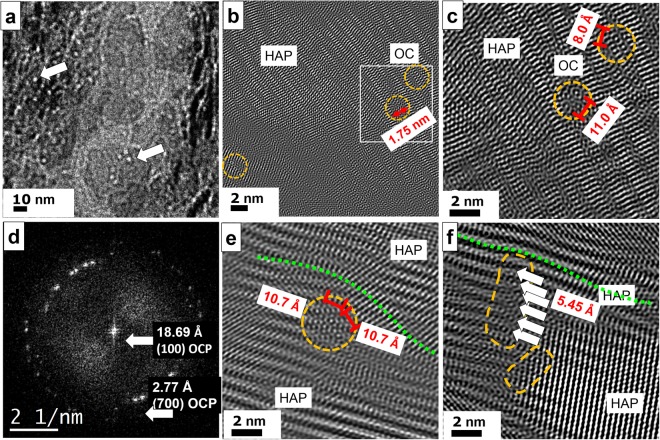
Figure 8.
TEM of conventional ultrathin diamond cuts of human femur. (a) Overview image with pearl bead strings of 2 nm diameter indicating the presence of osteocalcin, see arrows. At the center bottom a 50 × 50 nm2 large HAP crystal plate is imaged with OC particles on its surface resembling to white pearl bead structures suppressing the HAP growth normal to the plate plane. (b) The Fourier filtered high–resolution image displays a massive HAP crystal at the top of the micrograph. At certain regions, the crystal lattice is disturbed due to the presence of OC (see orange circles at right). (c) At higher magnification a gap within the crystal plate is revealed corresponding to the size of OC proteins. Even the binding sites are resolved which appear as small circles with pronounced dots in the center (distances of 11 and 8 Å). (d) The FFT of the high–resolution image show reflections at small angle corresponding to 18.69 Å, thus proving the presence of OCP. (e) OC is also found between HAP crystals with different orientations serving as delimiter. The insertion of OC evokes mechanical stress and leads to remarkable lattice distortions of the upper crystal (green dotted line). The binding sites within OC show 10.7 Å distance from each other. (f) The massive crystal plate on the right is confined by a string of OC particles marked in orange. At least six binding sites (see arrows) are arranged in a row.

