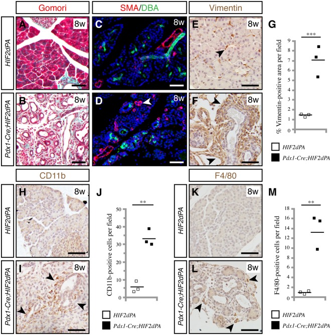
Figure 2.
HIF2α stabilization causes pancreatic abnormalities reminiscent of pancreatitis. Gomori trichome staining reveals increased connective tissue (stained in blue) in Pdx1-Cre;HIF2dPA pancreata (B) compared to control pancreata (A) at 8 weeks of age. Increase in smooth muscle actin (SMA) positive cells (white arrowhead) in Pdx1-Cre;HIF2dPA pancreata (D) compared to control pancreata (C) at 8 weeks of age. Ducts are marked by staining with lectin Dolichos biflorus agglutinin (DBA). Increase of the mesenchymal marker vimentin in Pdx1-Cre;HIF2dPA pancreata (F, black arrowheads) at 8 weeks of age. Only a few scattered cells are positive for vimentin in control pancreata (E, black arrowhead). (G) Quantification of vimentin-positive area per total pancreatic area. CD11b immunohistochemistry in control (H) and Pdx1-Cre;HIF2dPA pancreata (I, black arrowheads) at eight weeks of age. (J) Quantification of CD11b-positive cells per field. F4/80 immunohistochemistry in control (K) and Pdx1-Cre;HIF2dPA pancreata (L, black arrowheads) at eight weeks of age. (M) Quantification of F4/80-positive cells per field. Data points represent values for each individual mouse. The mean value is indicated as a horizontal line. Scale bars = 50 µm. **P < 0.01; ***P < 0.001.

