Figure 6.
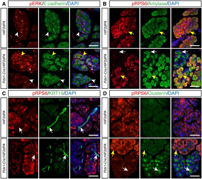
Altered components of MAPK and AKT/mTOR signaling pahtways in HIF2α-overexpressing pancreata. (A) Periacinar cells positive for phosphorylated-ERK (pERK) are found in 2-week-old control pancreata but acinar cells are negative for pERK (white arrowheads, top panels). Increased pERK accumulation is observed in 2-week-old Pdx1-Cre;HIF2dPA pancreata (yellow arrowheads, bottom panels). Note that the increased expression was limited to areas with acinar-ductal metaplasia (yellow arrowheads, bottom panels). Acini with preserved morphology do not show extensive pERK accumulation (white arrowheads, bottom panels). pERK-positive stromal cells are also observed in Pdx1-Cre;HIF2dPA pancreata (asterisks, bottom panels). E-cadherin immunofluorescence was performed to visualize the pancreatic epithelium. (B) Double immunofluorescence analysis of phosphorylated RSP6 (pRSP6) and amylase in 2-week-old mice. pRPS6 (yellow arrows) is detected in amylase-positive acinar cells of control pancreata (top panels). In Pdx1-Cre;HIF2dPA pancreata, pRPS6 is detected in well-preserved acini (bottom panels, yellows arrows). However, areas with acinar-ductal metaplasia display decreased expression of pRPS6 (white arrows). (C) Double immunofluorescence analysis of pRSP6 and KRT19 in 2-week-old mice. Ductal cells (white arrows) are not positive for pRPS6 in both control pancreata (top panels) and Pdx1-Cre;HIF2dPA pancreata (bottom panels). (D) Clusterin is not expressed in control pancreas (top panels). Areas with acinar-ductal metaplasia in Pdx1-Cre;HIF2dPA pancreata display decreased expression of pRPS6 and activation of clusterin expression (white arrows). Yellow arrows indicate acini with normal morphology. Nuclei are stained with DAPI (blue). Scale bars = 50 µm.

