FIG 3.
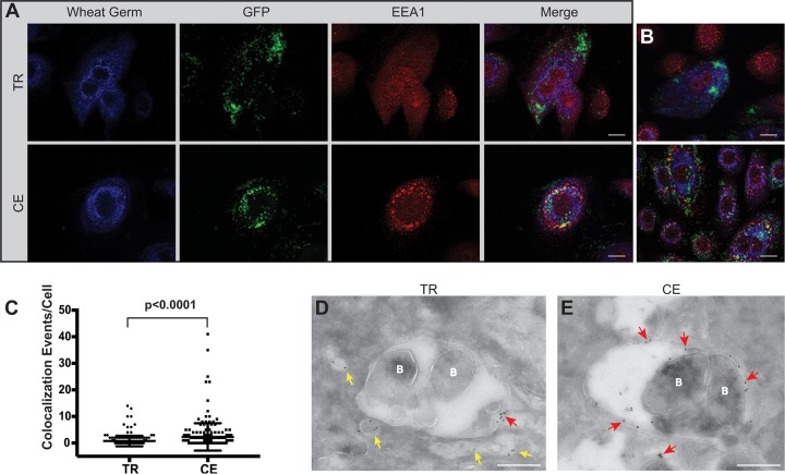
Transient heme-iron conditioning of NTHI alters trafficking to early endosomes. (A) NHBE cells were cocultured with either transiently restricted (TR) or continuously exposed (CE) NTHI strain 86-028NP(pGM1.1) for 4 h. Epithelial cell membranes were labeled with wheat germ agglutinin conjugated to Alexa Fluor 350 (blue), and bacteria were visualized by GFP fluorescence (green). Early endosomes were labeled with rabbit antibody to early endosomal antigen 1 (EEA1) protein and visualized with donkey anti-rabbit IgG conjugated to Alexa Fluor 594 (red). Representative images are shown for each condition (TR or CE) with individual and merged fluorescence images shown for depiction of colocalization. Colocalization of bacteria with EEA1 is observed as either yellow (merged) or red EEA1 label closely surrounding clusters of green NTHI bacteria. Bar, 10 µm. (B) Additional images representative of those depicted in panel A. (C) The number of colocalization events per cell was quantified by visual assessment of 200 individually infected cells. Statistical significance was determined by Mann-Whitney U test, and error bars represent standard errors of the means. (D and E) Transmission electron microscopy of NHBE cells cocultured with TR or CE conditioned NTHI for 4 h and subsequently immunolabeled to detect bacterial association with EEA1. Early endosomes were labeled with rabbit antibody to EEA1 and visualized with anti-rabbit IgG antibody conjugated to an 18-nm colloidal gold particle. EEA1-containing vesicles devoid of bacteria (labeled B) are indicated by yellow arrows, while red arrows indicate EEA1-containing vesicles associated with bacteria. Bar, 500 nm.

