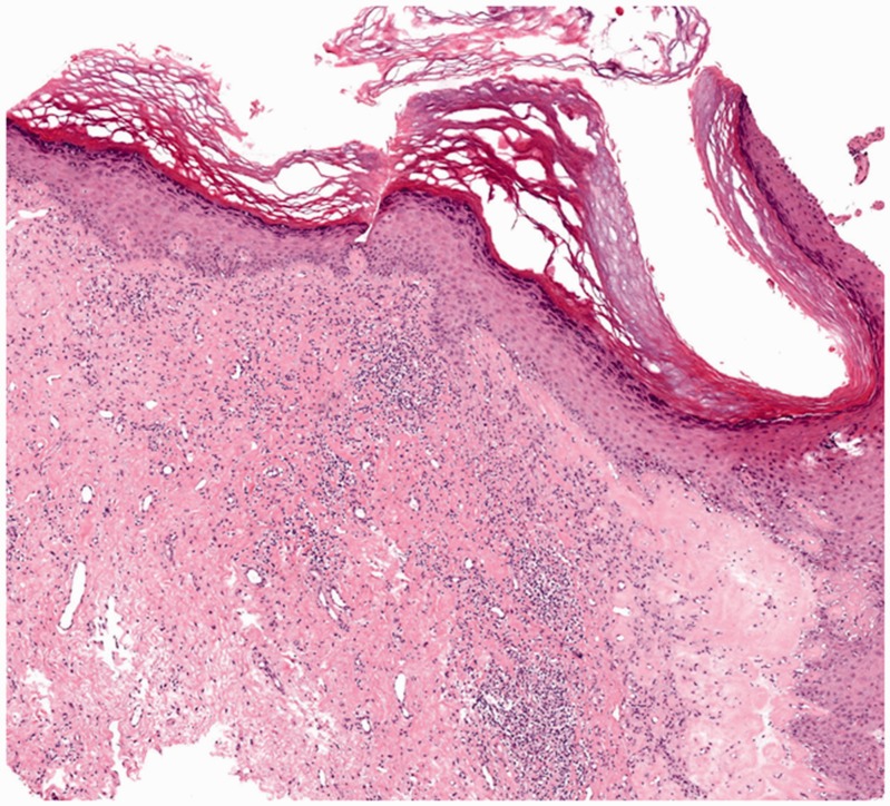
Figure 1.
Histological image of a patient with vulvar lichen sclerosus. Orthokeratotic hyperkeratosis of the epithelium and vacuolization of keratinocytes of the basal layer can be seen. Additionally, focal homogenization of the papillary dermis, perivascular, often band-like, infiltrate of lymphocytes, and plasma cells in the dermis can be seen (haematoxylin–eosin stain, ×40).

