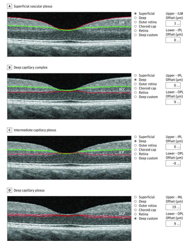
Figure 1. Locations of the Segmentation Lines Used to Determine Each Plexus.
The superficial vascular plexus (SVP) (A) and the deep capillary complex (DCC) (B) are automatically segmented. C, The intermediate capillary plexus (ICP) slab is obtained from the DCC by moving the outer boundary (red line) 9 µm above the outer plexiform layer–outer nuclear layer (OPL-ONL) junction. D, The deep capillary plexus (DCP) inner boundary is set 19 µm below the inner nuclear layer–outer plexiform layer (OPL) junction and the outer boundary is set 9 µm below the OPL–outer nuclear layer junction. ILM indicates internal limiting membrane; INL, inner nuclear layer; and IPL, inner plexiform layer.

