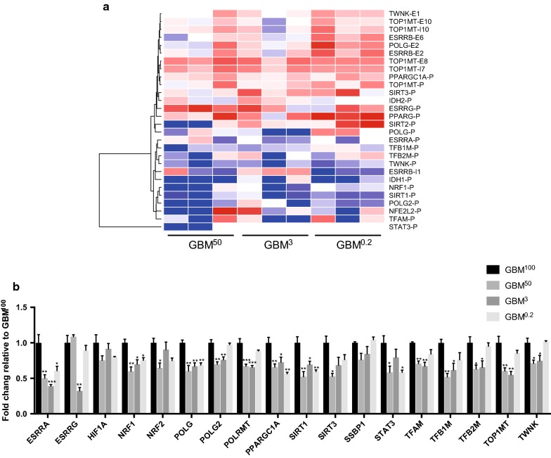
Fig. 6.
Overview of the changes to DNA methylation and transcription of the mtDNA replication factors. a DNA methylation levels of the intragenic CGIs associated with the mtDNA replication factors. Fold changes in relative methylation scores to the GBM100 tumours were plotted for the three biological replicates from the GBM50, GBM3 and GBM0.2 cohorts. The colour scheme from blue, white to red represents the level of DNA methylation from low to high. “P”, “E” and “I” following each gene name indicates CGIs that are located at the promoter regions, exons and introns, respectively. b Differential expression of the mtDNA replication factors. Bars represent the mean of the relative quantification levels relative to the GBM100 tumours (relative expression = 1). Error bars show SEM (n = 3 tumours per group; technical replicates per sample). Statistical significance was determined by one-way ANOVA. *, ** and *** p values < 0.05, 0.01 and 0.001, respectively

