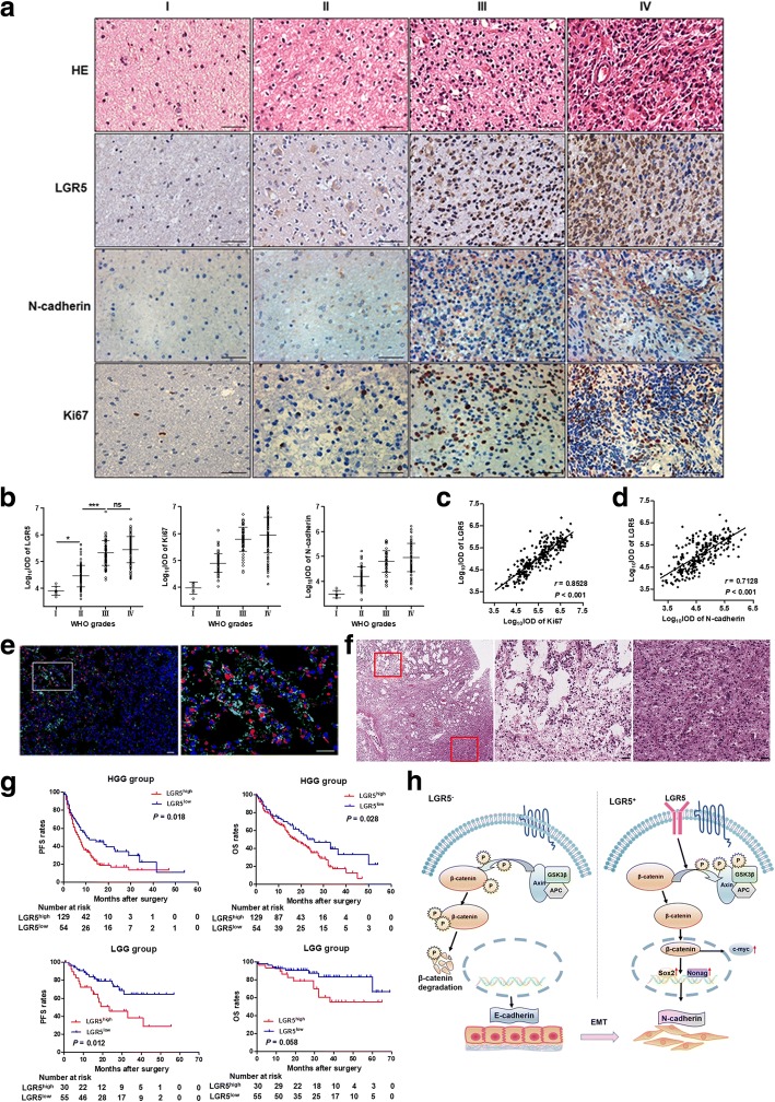
Fig. 6.
Immunohistochemistry analyses of human glioma specimens. a HE and LGR5, Ki67 and N-cadherin staining of human glioma specimens from four patients in four WHO grades. Scale bar = 100 μm. b Quantitative analyses of LGR5, Ki67 and N-cadherin in four WHO grades (WHO I and II, n = 85; WHO III and IV, n = 183, Student t test). Data are showed as mean ± SD. c Spearman correlation analyses of LGR5 and Ki67 (P < 0.001, r = 0.8528, n = 268). d Spearman correlation analyses of LGR5 and N-cadherin (P < 0.001, r = 0.7128, n = 268). e LGR5 and N-cadherin staining in glioma tissues. Many glioma cells with high expression of LGR5 and N-cadherin were mostly observed in the invasive edge of tumor (left of 1st picture) compared with the interior of tumor (right of 1st picture). Scale bar = 100 μm. f The HE staining of the glioma tissues. The structure of tumor edge (left top), consistent with the invasive edge of Fig. 6e, were more incompact compared with the inside tumor (right bottom). g PFS and OS months in low-grade glioma (LGG) group and high-grade glioma (HGG) group according to LGR5 expression in the glioma specimens. ***, P < 0.001; *, P < 0.05; ns, not significant. h A schema diagram displaying the role of LGR5 in regulating Wnt, and EMT in glioma stem cells. Based on the findings of this study, LGR5 could activate Wnt pathway by promoting β-catenin dephosphorylation and result in EMT eventually

