. 2018 Jul 23;41(3):570–577. doi: 10.1590/1678-4685-GMB-2016-0287

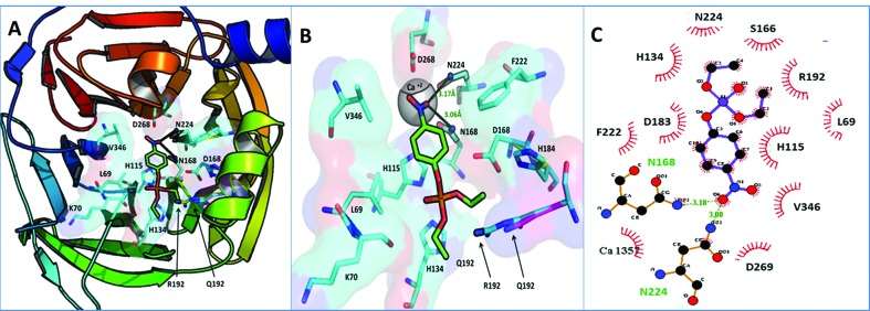
Figure 2. Docked conformations of paraoxon in PON1 binding pocket. (A) Paraoxon bound in PON1 binding pocket. (B) Zoomed in binding pocket of PON1, paraoxon terminally making two hydrogen bonds with N168 and N224. Notice that R192 adopts a conformation more proximal to the binding pocket opening than that of Q192, steering the ligand to adopt the most suitable conformation for paraoxonase activity by H 115 and H134 dyad. (C) 2D illustration of paraoxon binding in PON1 binding pocket; paraoxon forms two hydrogen bonds with Asn 224 (3.00 Å) and Asn 168 (3.18 Å) shown in green while a number of hydrophobic interactions with the residues configure the binding pocket.

Figure 2