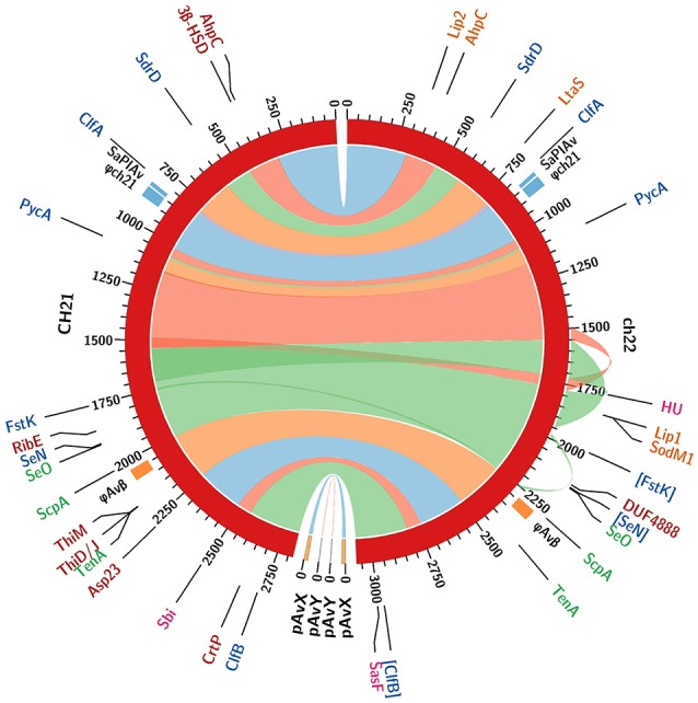
Figure 5.

Comparison of whole genome sequences of the CH21 and ch22 strains. Color bands represent continuous similar sequence segments characterizing the two strains. Particular segments are separated by short insertions or duplications unless indicated otherwise. Three longer duplications in the ch22 genome (170, 54, and 11 kbp) are additionally marked by outward ribbons. Loci of intrachromosomal mobile genetic elements, such as prophage φAvβ, putative prophage φch21 and pathogenicity island SaPIAv, are marked. The loci differentiating the two strains are identified with acronyms and colored as follows. Genomics: coding sequences in blue and promoters in green (pseudogenes in square brackets). Proteomics: intracellular in red; extracellular in orange; and surface in magenta (upregulated proteins only).
