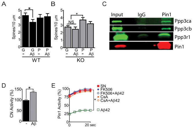
Fig. 3. Calcineurin interacts with Pin1 and mediates Aβ42 signaling.
(A and B) Total spine counts for DIV21 wild-type (WT; A) or DIV21 KO (B) neurons transduced with TAT-GFP (“G”) or TAT-Pin1 (“P”) ± Aβ42 (Aβ). Data are mean ± SEM; N >100 spines from ≥15 images and ≥ 3 coverslips per condition were counted. *P<0.05 by Fisher’s LSD test following two-way analysis of variance (ANOVA). (C) Western blot of atalytic and regulatory subunits of calcineurin inPin1 immunoprecipitations from synaptoneurosomes prepared from P28 mice. (D) Calcineurin (CN) activity in synaptoneurosomes that were either untreated (−) or treated with Aβ42 (Aβ) for 10 min before lysis. Data are mean ± SEM; N = 3 biological replicates. *P < 0.05 by an unpaired t-test. (E) Pin1 activity assay in synaptoneurosomes that were either untreated (SN) or treated with Aβ42, FK506, FK506 + Aβ42, CsA, or CsA + Aβ42 before lysis. Data are means from N ≥ 8 replicates per treatment. *P < 0.05 by Fisher’s LSD test following two-way analysis of variance (ANOVA).

