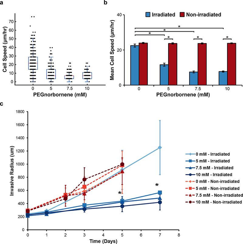
Figure 4: Analysis of Cell Motility within PEG/Collagen IPNs.

a) Box-whisker plot of migration speed of individual MDA.MB.231 cells embedded diffusely within PEG/Collagen IPNs over the course of 24 hours immediately following crosslinking. b) The average cell speed for the entire population of cells within the irradiated PEG/Collagen IPNs or non-irradiated controls (mean ± SEM; n = 3 gels, 2 locations per gel). P value determined using ANOVA with Tukey-Kramer post hoc analysis (*P < 0.001). c) The average invasive radius of MDA.MB.231 multicellular spheroids embedded within PEG/Collagen IPNs were measured as a function of time (mean ± SEM; n = 3 spheroids).
