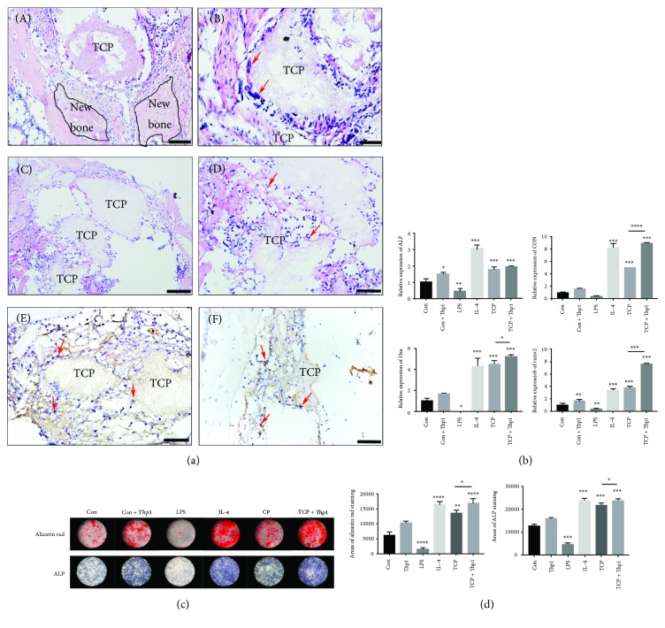
Figure 1.
Osteogenic effect and gathering of macrophages at the healing defects filled with TCP. (a) (A–D) Representative sections of hematoxylin-eosin staining. Macrophages are indicated by red arrows. (E) Representative sections of immunohistochemistry staining with F4/80 antibody. Macrophages are indicated by red arrows. (F) Representative sections of immunohistochemistry staining with CD206 antibody. M2 macrophages are indicated by red arrows. (A,C) Scale bar = 50 μm; (B, D, E, and F) scale bar = 20 μm. (b) Relative mRNA expression of ALP, OCN, OSX, and Runx2 of HBMSC stimulated by control medium, supernatant of control Thp1, LPS, IL-4, and TCP extract and supernatant of TCP-induced Thp1. (c) Alizarin red staining, ALP staining, and quantification of HBMSC stimulated by control medium, supernatant of control Thp1, LPS, IL-4, and TCP extract and supernatant of TCP-induced Thp1. (d) Quantification of the areas of alizarin red staining and ALP staining was measured by ImageJ software. ∗p < 0.05, ∗∗p < 0.01, ∗∗∗p < 0.001, and ∗∗∗∗p < 0.0001.

