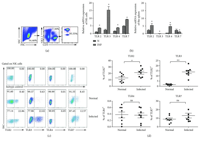
Figure 2.
Expression of different TLRs in mouse splenic lymphocytes and NK cells after S. japonicum infection. (a) Splenic lymphocytes were separated from normal and S. japonicum-infected wild-type mice, and CD3−NK1.1+ NK cells were isolated from splenic lymphocytes by using flow cytometry and the purity of isolated splenic NK cells was identified by FACS. (b) Total RNA of splenic lymphocytes and NK cells was harvested, respectively. The accumulation of TLR2, TLR3, TLR4, and TLR7 mRNA was quantified by using qPCR. The levels of TLR transcripts were normalized to β-actin transcripts by using the relative quantity (RQ) = 2−△△Ct method. Data represent means ± SEM of at least three experiments. (c, d) Expression of TLR2, TLR3, TLR4, and TLR7 on splenic NK cells was assessed by using flow cytometry. (c) A representative result is shown. (d) Statistic results of 6 to 8 independent results are shown. N: normal; INF: infected; ∗P < 0.05, comparison between the normal and infected groups.

