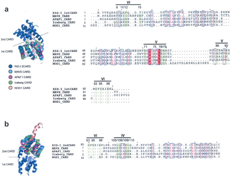
Extended Data Figure 9. Sequence analysis of the Ub-binding surface in the CARD family.
Structure-based sequence alignment (using the program SALGIN30) of various CARD domains. In our effort to further analyse potential generality of 2CARD–Ub interaction observed with RIG-I in our structure, we aligned other CARDs with RIG-I 2CARD. We performed structure-based sequence alignment, as many members of the CARD family share little sequence similarity. Three dimensional protein structure, which is more conserved than the primary sequence, allows more accurate sequence comparison. The first CARD (a) and second CARD (b) of human RIG-I was aligned with other CARDs from MAVS, APAF1, Iceberg and NOD1 (PDB code: 2VGQ, 3YGS, 1DGN and 4E9M, respectively). None of the Ub-binding residues in RIG-I 2CARD are conserved in these CARDs.

