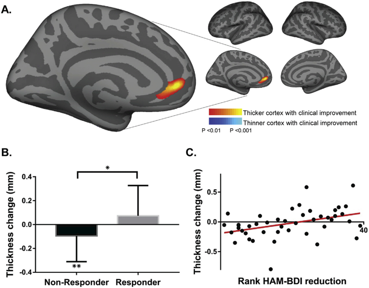
Fig. 1. Cortical thickness change correlates with clinical improvement.
A. A cortex-wide analysis revealed a significant correlation between cortical thickness change in left rostral anterior cingulate cortex (rACC) and improvement in depression symptoms. The peak correlation is significant at P < 0.001 uncorrected. Findings are shown at P < 0.01 to display the extent of the spatial distribution of the correlation, also see Fig. S3 showing these results at a P < 0.001 threshold. B. Average cortical thickness changes within this rACC region differ between responders and non-responders (+0.074 and −0.095 mm, respectively; P < 0.05) with non-responders showing a significant reduction in cortical thickness (P < 0.01). C. A scatter plot shows the correlation between change in cortical thickness within this ROI and clinical response (r = 0.4, P < 0.01). *P < 0.05, **P < 0.01.

