Fig 2.
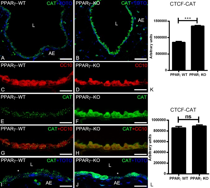
Immunofluorescence detection of the peroxisomal marker enzyme catalase in paraffin-embedded lung tissue sections of wild-type (2A, 2C, 2E, 2G, 2I) and PPARγ-deficient club cells (2B, 2D, 2F, 2H, 2J). Note that, PPARγ-deficiency increased the catalase protein abundance. Moreover, the catalase is partially mistargeted into the cytoplasm (2F, 2H, 2J). Club cells-specific protein CC10 stained club cells (2C, 2D) and higher magnifications (2E and 2F) stained for catalase showing the cross sections of terminal bronchioles of the mouse lung. Cytoplasmic staining of catalase was observed in CC10 positive club cells (2G, 2H). Staining for catalase showed punctuate staining pattern in higher amounts in club cells in comparison to ciliated cells (*) in WT animals, whereas it is present in cytoplasm (in addition to peroxisomes) in ccsPPARγKO club cells, but neighboring ciliated cells still contain punctuate catalase with similar amounts (2I, 2J, 2K, 2l). Corrected total cell fluorescence (CTCF) quantification of staining for CAT in club cells (2K) and ciliated cells (2I). Values ± SEM represent the mean of CTCF quantified from images obtained from 3 independent experiments using Image J software. **P ≤0.01; ***P ≤0.001; ns, not significant. Counterstaining of nuclei was performed with TOTO-3 iodide in 2A, 2B, 2I, 2J. 2L: lumen of the bronchiole; AE: alveolar epithelium. *: ciliated cell; Bars represent 2A-2B: 100 μm; 2C-2H: 25 μm.

