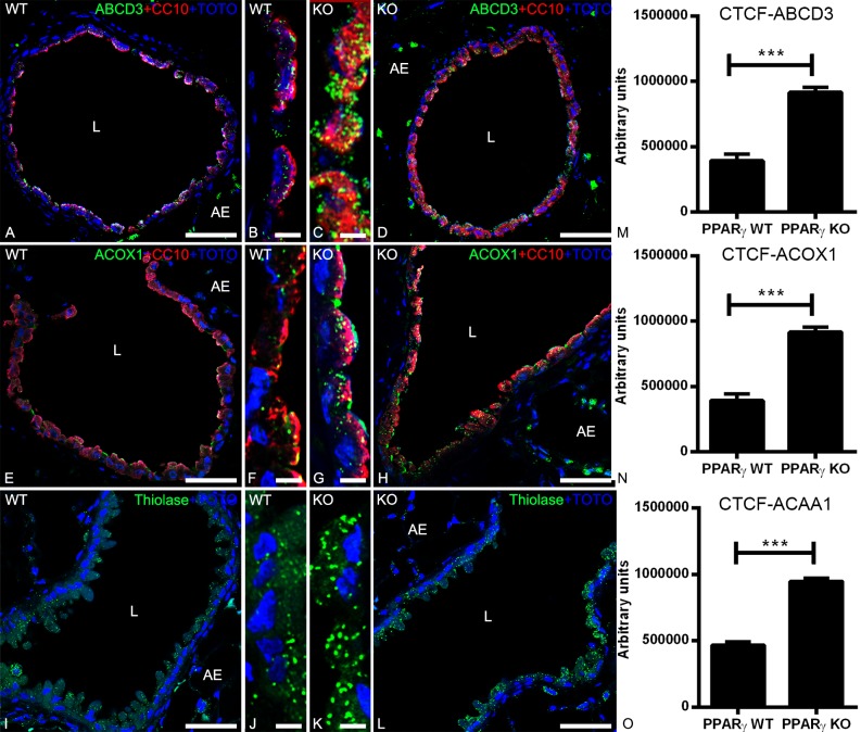
Fig 3. Double-immunofluorescence detection of peroxisomal proteins together with CC10 in the bronchioles of WT and PPARγ-deficient club cells.
Staining of mouse lung tissue sections of WT and ccsPPARγKO for peroxisomal lipid transporter ABCD3 (3A-3D) or peroxisomal β-oxidation enzymes ACOX1 (3E-3H) and thiolase (3I-3L). Note that ABCD3 (3C-3D), ACOX1 (3G-3H) and thiolase (3K-3L) protein abundance is increased in PPARγ-deficient club cells. Representative lower (3A, 3D, 3E, 3H, 3I, 3L) and higher magnifications (3B, 3C, 3F, 3G, 3J, 3K) of cross sections of bronchioles in the mouse lung are depicted. Corrected total cell fluorescence (CTCF) quantification of staining for ABCD3 (3M), ACOX1 (3N), and thiolase (3O) in club cells. Values ± SEM represent the mean of CTCF quantified from images obtained from 3 independent experiments using Image J software. **P ≤0.01; ***P ≤0.001; ns, not significant. Counterstaining of nuclei was performed with TOTO-3 iodide. 3L: lumen of the bronchiole; AE: alveolar epithelium. Bars represent: 3A, 3D, 3E, 3H, 3I and 3L: 50 μm; 3B, 3C, 3F, 3G, 3J and 3K: 5 μm.

