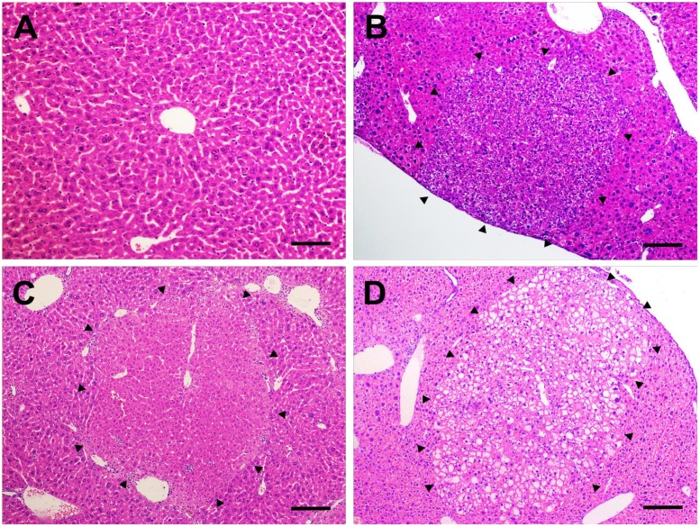
Fig 3. Preneoplastic altered hepatocyte foci.
Representative photomicrographs of (A) normal liver of vehicle-treated groups (20× objective) (scale bar = 50 μm) and (B) basophilic, (C) eosinophilic and (D) clear cell preneoplastic foci found in the liver of DEN/CCl4-treated mice (10× objective) (scale bar = 100 μm).

