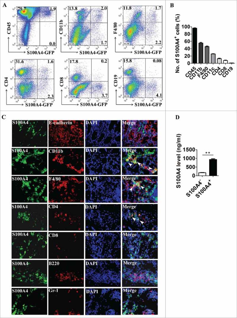
Figure 2.

S100A4 is expressed in different types of cells in the colon. (A-B) Flow cytometry analysis of the phenotypes of S100A4+ cells in the colons of S100A4+/+.GFP mice treated with 3% DSS for 5 days for 2 cycles by staining GFP+ cells with CD45, CD11b, F4/80, CD11 c, CD4, CD8 and CD19 antibodies. (C) Double immunohistochemical staining of S100A4 with E-cadherin, CD11b, F4/80, CD4, CD8, B220 and Gr-1 in DSS-induced colon tissues. Nuclei were counter-stained with DAPI. The arrows indicate double-positive cells. Scale bar, 25 μm. (D) S100A4 concentrations in the cultured supernatants of S100A4+ CD11b+ cells or S100A4− CD11b+ cells as detected by ELISA. **P < 0.01.
