Figure 4.
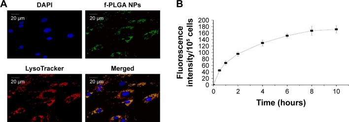
Intracellular distribution and internalization kinetics of f-PLGA NPs in MSCs.
Notes: (A) Confocal laser-scanning microscopy of MSCs treated with f-PLGA NPs (5 µg/mL). Lysosomes and nuclei were stained with LysoTracker Red and DAPI, respectively. (B) Uptake kinetics of f-PLGA NPs into MSCs.
Abbreviations: f-PLGA NPs, fluorescein–poly(d,l-lactide-co-glycolide) nanoparticles; MSCs, mesenchymal stem cells.

