
Figure 9.
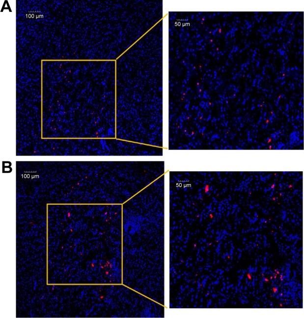
Intracerebral distribution of MSC NPs.
Notes: (A) Confocal laser-scanning microscopy of glioma. All the area was within the glioma. (B) Brain tissue at the therapeutic injection site 2 days after contralateral injection of MSC NPs. MSCs were incubated with 8 ng/mL Ptx-PLGA NPs for 8 hours, and labeled with a red fluorescent probe – CM-Dil. Nuclei were stained with DAPI before observation.
Abbreviations: Ptx-PLGA NPs, paclitaxel poly(d,l-lactide-co-glycolide) nanoparticles; MSC, mesenchymal stem cell.