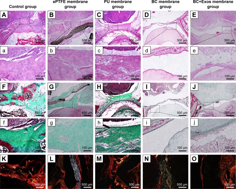
Figure 7.
Histological examination.
Notes: (A, a, B, b, C, c, D, d, E, and e) The HE staining images of the control, ePTFE membrane, PU membrane, BC membrane, and BC+Exos membrane groups at 1 year post operation, respectively. (F, f, G, g, H, h, I, i, J, and j) The Masson trichrome images of the control, ePTFE membrane, PU membrane, BC membrane, and BC+Exos membrane groups at 1 year post operation, respectively. (K–O) The Sirius red staining of the control, ePTFE membrane, PU membrane, BC membrane, and BC+Exos membrane groups at 1 year post operation, respectively.
Abbreviations: BC, bacterial cellulose; BC+Exos, bacterial cellulose combined with exosomes; ePTFE, expanded polytetrafluoroethylene; MRI, magnetic resonance image; PU, polyurethane.

