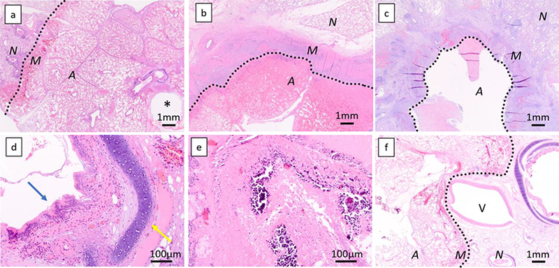
Figure 3. Histology of ablation zone.

Histological specimen at day 2 (a, 1,25x, short duration) shows a well demarcated area of complete coagulation necrosis with a rim of hyperemia, hemorrhage, and fibrin exudation at the edge of the area of necrosis. At day 28, a rim of dense fibrosis and inflammation characterized by infiltration by numerous neutrophils, macrophages, lymphocytes, and plasma cells is seen (b, 1.25x, long duration). Some lesions show cavitation within the dense rim (c, 1.25x, short duration). All bronchi were completely ablated inside the ablation zone. At day 2, the structure, including epithelium (blue arrow) or cartilage (yellow arrow) of bronchus is maintained (d, 20x, short duration energy delivery), but is destroyed at day 28 (e, 20x, short duration energy delivery). A 5mm pulmonary artery at the edge of the necrotic zone shows incomplete necrosis, and the ablation zone margin is concaved (f, 1.25x, short duration energy delivery). A, ablated area; M, ablation margin; N, non-ablated area; *, antenna tract; V, pulmonary vein. Dotted lines demarcate the ablation zone from untreated lung.
