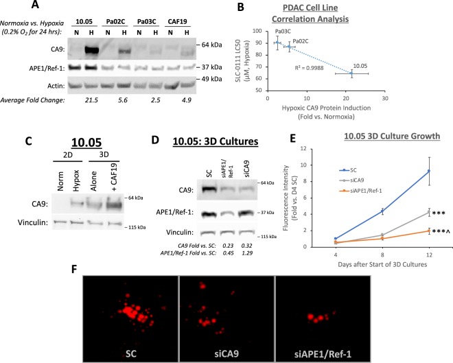
Figure 1.
Hypoxia-induced CA9 expression and constitutive APE1/Ref-1 expression are important for 3D PDAC tumor spheroid growth. (A) Patient-derived PDAC tumor cell lines 10.05, Pa02C, and Pa03C, as well as the pancreatic CAF cell line CAF19, were exposed to 0.2% oxygen for 24 h, and CA9 protein levels were compared via western blot (p < 0.05 for all cell line differences between normoxia and hypoxia). Representative blot of N = 3. (B) LC50 values for SLC-0111 in PDAC cell lines under hypoxic conditions (0.2% O2) are inversely correlated with CA9 induction in each cell line (R2 > 0.99, N = 3). (C) Comparison of CA9 protein induction between 10.05 cells grown in monolayer (2D, 0.2% O2, 24 h) and in 3D cultures alone or in co-culture with CAF19 cells for 12 days. N = 1 (D–F). 10.05 Cells were transfected with the indicated siRNAs and cultured in 3D spheroids. Cells were collected for western blot analysis on Day 8 to confirm knock-down (D, p < 0.05 for siAPE1/Ref-1 and siCA9 effects on CA9 as well as siAPE1/Ref-1 effects on APE1/Ref-1, N = 3). Fluorescence intensity (E) of both tumor (TdTomato) and CAFs (EGFP) was measured to track 3D tumor growth on days 4, 8, and 12 of 3D cultures (+/−SEM, ***p < 0.001 for differences between knockdown groups and SC on D12, ^p < 0.05 for difference between siAPE1/Ref-1 and siCA9 on D12, N = 3). Representative fluorescent images from each group were captured on day 12 (F). Full size blots are shown in Supplemental Figs S3–S5.

