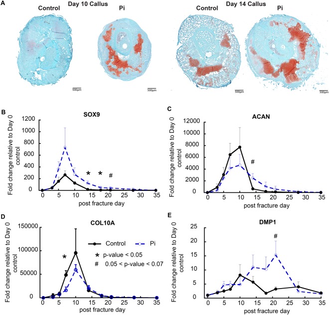
Figure 2.
Effects of hypophosphatemia on the progression of cartilage and bone cell differentiation and identification of altered circadian function during fracture healing. (A) Histological analyses of the effects of hypophosphatemia on post-operative day 10 and day 14 callus tissues. Callus sections in the mid-transverse plane of control and hypophosphatemic (Pi) mice are shown. Callus tissues from hypophosphatemic mice displayed large regions of residual cartilage and large regions of lesser differentiated cells tissue abutting the fracture. (B–E) Expression of cartilage and bone-related genes as denoted in the figure: Error bars represent ±1 standard error of the mean of the 2–3 repeat analyses performed on six samples per group. *A significant t-test p-value comparing Pi and Control expression levels at each time point (p < 0.05). #A trend in t-test p-value comparing Pi and Control expression levels at each time point (0.05 < p < 0.07).

