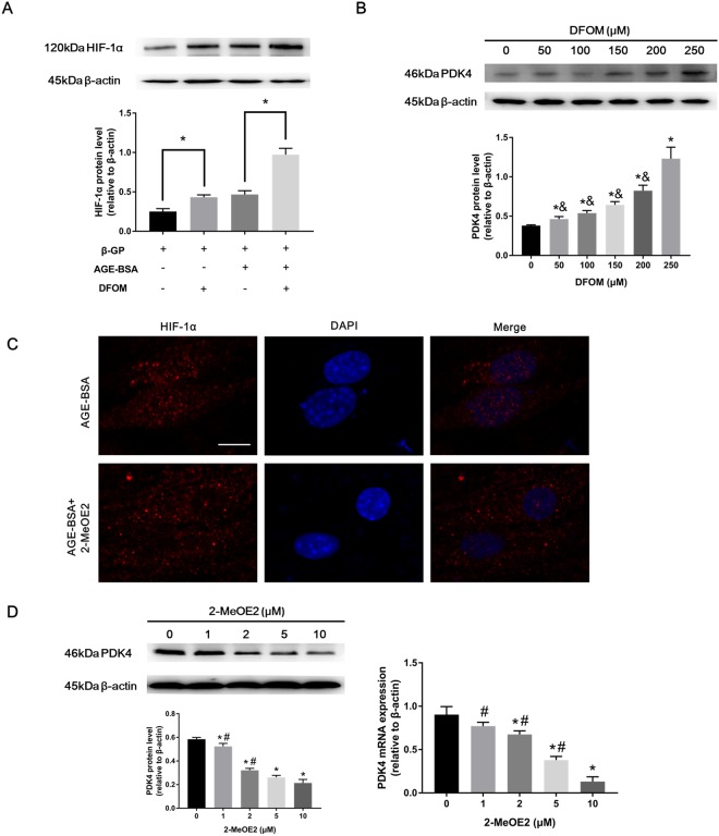
Figure 4.
PDK4 is associated with HIF-1α during VSMC calcification. (A) Calcified VSMCs were pretreated with DFOM (250 μM) for 6 h and then cultured with or without AGE-BSA (200 μg/ml) for 24 h. HIF-1α protein levels were determined by western blotting. *P < 0.05 vs. the indicated treatment. (B) Calcified VSMCs were preincubated with DFOM for 6 h, and the cells were exposed to AGE-BSA (200 μg/ml) for another 24 h. PDK4 expression was detected by western blotting. *P < 0.05 compared with the normal control group. &P < 0.05 compared with the DFOM (250 μM) group. (C) Calcified VSMCs were pretreated with 2-MeOE2 (10 μM) for 2 h and then incubated with or without AGE-BSA (200 μg/ml) for 24 h. HIF-1α nuclear translocation in VSMCs was visualized by immunofluorescence staining; scale bar: 10 μm (D) After 2 h of 2-MeOE2 exposure, calcified VSMCs were incubated as indicated. PDK4 expression was detected by western blotting and qRT-PCR. *P < 0.05 compared with the normal control group. #P < 0.05 compared with the 2-MeOE2 (10 μM) group.

