FIGURE 6.

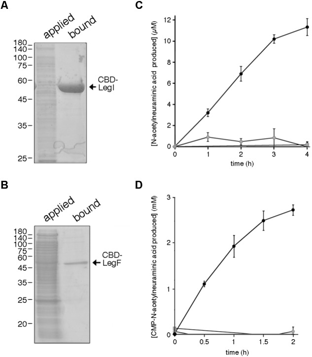
Halorubrum sp. PV6 LegI and LegF contribute to legionaminic acid biosynthesis. (A) Halorubrum sp. PV6 LegI and (B) LegF were purified from total lysates of Hfx. volcanii expressing each CBD-tagged protein (applied) on cellulose (bound). Aliquots of each pool were separated by 12% SDS-PAGE and Coomassie stained. In each panel, the positions of molecular weight markers are indicated on the left, while the position of the purified protein is indicated on the right. (C) LegI activity was confirmed in reactions containing cellulose-bound CBD-LegI, N-acetylmannosamine, and phosphoenolpyruvate (black circles) or in which the N-acetylmannosamine (gray circles) or phosphoenolpyruvate (gray squares) were omitted, or where cellulose incubated with the lysate of Hfx. volcanii not expressing the CBD-tagged protein was added instead of CBD-LegI-bearing beads (gray triangles). Each point is the average of three repeats ± SEM. (D) LegF activity was confirmed in reactions containing cellulose-bound CBD-LegF, N-acetylneuraminic acid and CTP (black circles) or in which the N-acetylneuraminic acid (gray circles) or CTP (gray squares) were omitted, or where cellulose incubated with the lysate of Hfx. volcanii not expressing the CBD-tagged protein was added instead of CBD-LegF-bearing beads (gray triangles). The amount of N-acetylneuraminic acid produced in each reaction was determined every 30 min over a 2 h interval, except in the latter two controls, when measurements were only taken at the start and the end of the experiment. Each point is the average of three repeats ± SEM.
