Fig. 2.
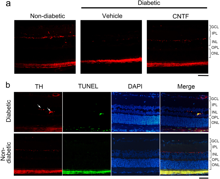
Immunohistochemistry of TH and TUNEL staining on paraffin sections of rat retinas. a In the rat retinas, the TH-positive amacrine neurons and fibers were observed in the innermost row of the INL. TH-positive fibers of the IPL could be detected in the non-diabetic rat retina. In the diabetic animals, TH-positive fibers in the IPL cannot be detected, and the fibers in the INL were thinner than in non-diabetic animals. CNTF can reduce the loss of TH-positive cells and fibers in diabetic animal retinas. b TUNEL staining was carried out in combination with immunostaining against TH. In the images, the TH-positive neurons (red) showed a TUNEL-positive signal (green), confirming that apoptosis occurs in the dopaminergic amacrine cells of diabetic animals. No apoptosis event was detected in the retinas of non-diabetic animals. Scale bar = 50 μm

