Figure 1.

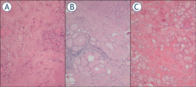
Haematoxylin and eosin stained sections of Case 4 (A), Case 5 (B) and Case 6 (C). Follicular atrophy and fibrosis, fibrosis accompanied by chronic inflammatory cells and fibrosis are seen, respectively.

Haematoxylin and eosin stained sections of Case 4 (A), Case 5 (B) and Case 6 (C). Follicular atrophy and fibrosis, fibrosis accompanied by chronic inflammatory cells and fibrosis are seen, respectively.