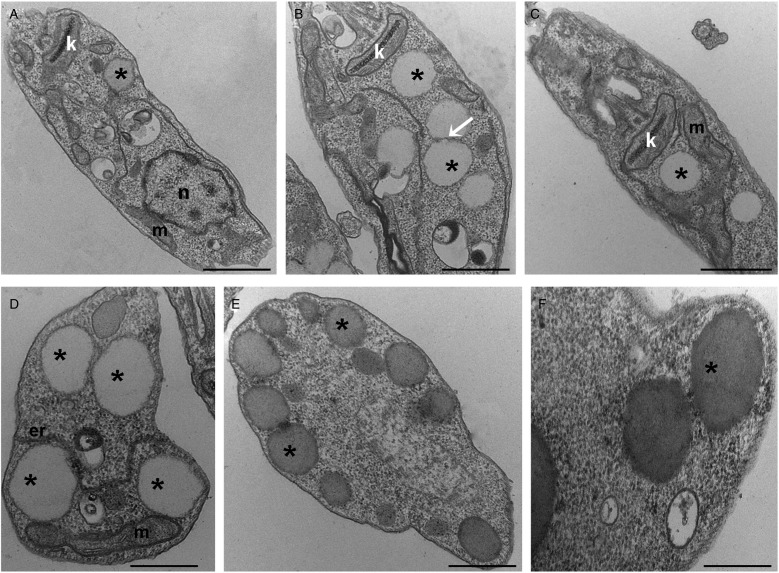
Fig. 2.
Transmission electron microscopy ultrastructural analysis of Leishmania amazonensis promastigotes treated with lopinavir. Untreated parasites (A) or those treated with ½IC50 (B), IC50 (C and D) and 2 × IC50 (E and F) of lopinavir for 72 h are shown. Lipid inclusions (asterisks) are numerous in parasites treated with lopinavir (B–H). Sometimes it is possible to detect fusion between lipid bodies (B – arrow). Such structures were observed in close association with the mitochondrion (C and D) and the endoplasmic reticulum (D). Lipid bodies are commonly observed at the cell periphery (E), close to the plasma membrane and even promoting its protrusion (F). M – mitochondrion, N – nucleus, ER – endoplasmic reticulum. Bars equal to 2 µm (A–C), 1 µm (D and E) and 0.5 µm (F).

