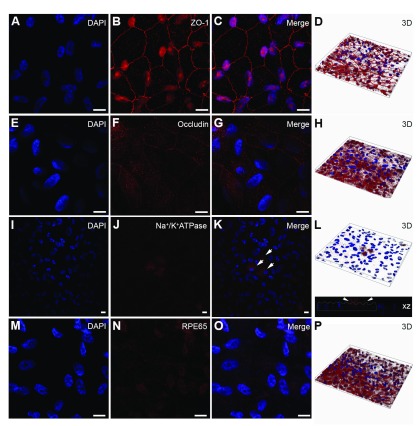
Figure 4. Characterisation of ARPE-19 monolayers cultured on transwell inserts.
Cultures were probed for [ A– D] the early tight junctional marker Zonula Occludens-1, which showed cobblestone morphology characteristic of Retinal Pigment Epithelial (RPE) cells. [ E– H] We also observed expression of the mid-late barrier protein occludin, although their staining was somewhat weaker. [ I– L] The epithelial transporter Na +/K + ATPase was observed in APPE-19 monolayers (arrows) but was not evident in all cells. Staining was however observed predominantly on the apical RPE surface, a feature reported in highly differentiated RPE cells. [ M– P] The cell-specific marker RPE-specific 65 kDa protein (RPE65) was also observed after 2 months in culture. Nuclei were counterstained with DAPI (blue). [ A– C, E– G, I– K, M– O] show representative en-face confocal images whilst [ D, H, L, P] show corresponding z-plane reconstructions. Scale bars correspond to 20μm.

