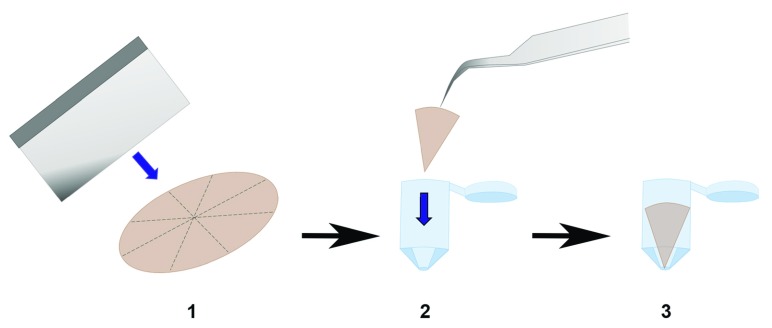
Figure 5. Preparation of polyethylene terephthalate (PET) membranes for transmission electron microscopy (TEM).
Schematic outlining steps carried out to embed segmented transwell membranes into capsules containing fresh Spurr resin. The apex is positioned downwards, which greatly assists with cutting sections for TEM.

