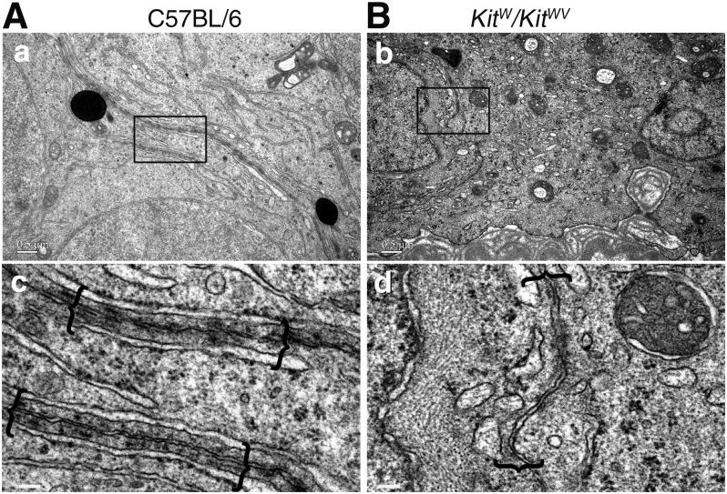
Figure 2.
Structural status study of KitW/KitWV testes by electron microscopy. A) Electron micrographs illustrating presence of typical BTB ultrastructure (enclosed in brackets) in control C57BL/6 mice (a, c). B) No typical BTB ultrastructures were detected in seminiferous epithelium of KitW/KitWV mouse testes without germ cells; only some irregularly shaped vesicles were noted on inner sides of adjacent Sertoli cell membranes (b, d). Boxed areas were magnified and are shown in lower panel. Scale bars, 0.5 μm (a, b), 100 nm (c, d).

