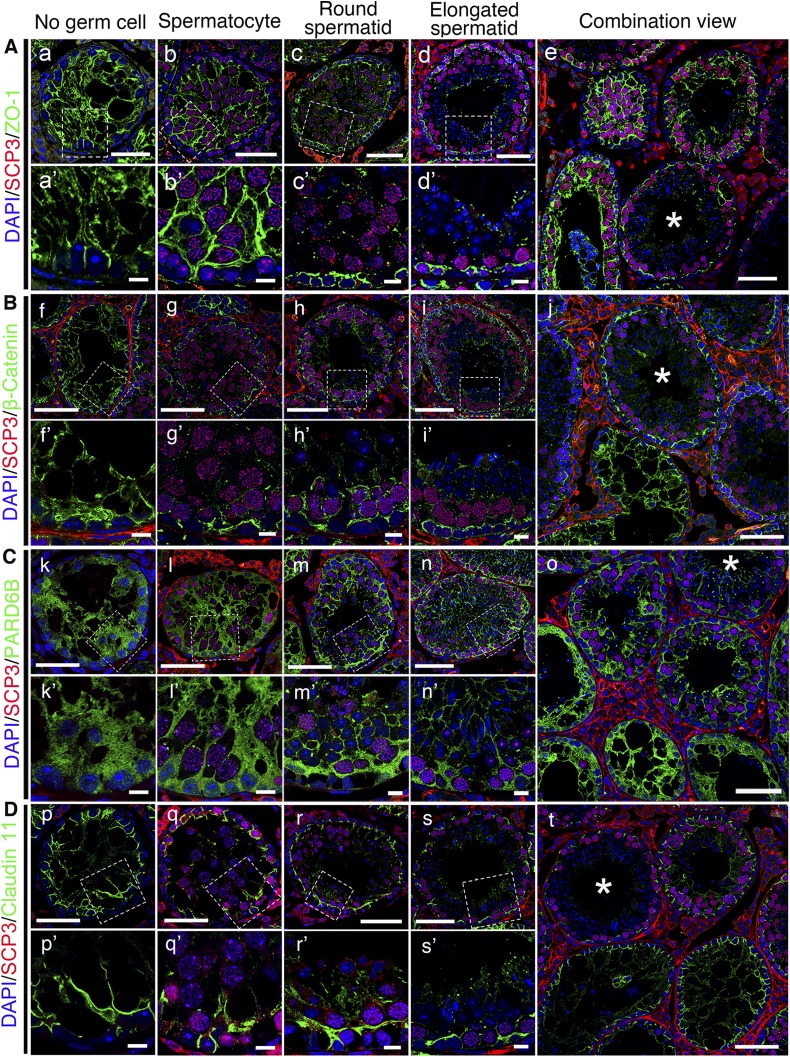
Figure 3.
Changes in distribution of BTB constituent proteins after SSC transplantation, which is dependent on status of germ cell differentiation and spermatogenesis. Distribution of ZO-1, β-catenin, PARD6B, and claudin 11 (all green) were found to be different in different tubules, depending on status of spermatogenesis and differentiation status of germ cells. Abnormal distribution of these proteins in empty tubules devoid of germ cells (a, f, k, p) was obviously noted compared to tubules that gradually returned to normal after appearance of spermatocytes (b, g, l, q) and status of spermatogenesis was fully restored when round spermatid appeared (c, h, m, r). Spermatocytes were labeled by anti-SCP3 antibody (red); round spermatids and elongated spermatids were identified by corresponding nucleus shape (blue). Tubules with complete spermatogenesis are marked with an asterisk in e, j, o, t. Boxed areas in a–d, f–i, k–n, p–s are magnified and shown in a′–d′, f′–i′, k′–n′, p′–s′. Scale bars, 50 μm (a–t), 10 μm (a′–s′).

