Figure 4.
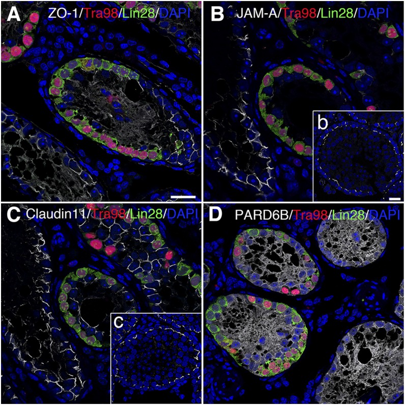
Changes in expression pattern of BTB constituent proteins in seminiferous epithelium after mutant SSC transplantation. Mutant SSCs were capable of homing in on basement membrane and proliferated such that primitive germ cells encircle base of seminiferous tubules. Germ cells marked by Tra98 (red) were positive for spermatogonia marker Lin28 (green). A–C) Signals (silver-gray) of ZO-1, JAM-A, and claudin 11 between adjacent Sertoli cells disappeared after SSC transplantation. No right position pattern as shown in b, c could be seen in recipient testes (normal position pattern of ZO-1 and PARD6B shown in Fig. 1B). D) PARD6B was diffusely localized both in tubules with and without SSCs. Scale bars, 20 μm.

