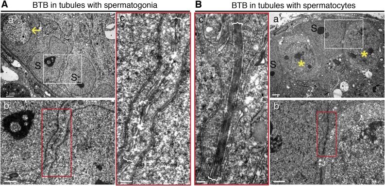
Figure 5.
Electron micrographs to confirm presence of BTB in seminiferous epithelium with spermatocytes vs. spermatogonia. A) Electron micrograph showed no typical BTB ultrastructure in tubules where only undifferentiated spermatogonia (yellow arrows) homed in on mouse testis after transplantation of mutant SSCs, displaying discontinuous and apparently truncated cell junctions. B) Typical BTB ultrastructures were detected in tubules where SSCs had differentiated into spermatocytes (yellow asterisk) in mice where normal SSCs had been transplanted. S, Sertoli cells. Boxed areas were magnified and are shown in lower or lateral panel. Scale bars, 2 μm (a, a′), 1 μm (b, b′), 0.4 μm (c, c′).

