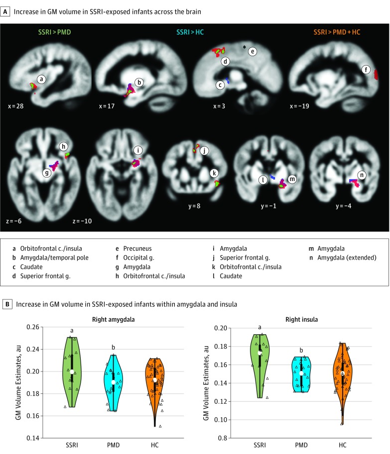
Figure 1. Brain Region Volumes in Infants With Prenatal Selective Serotonin Reuptake Inhibitor (SSRI) Exposure.
A, Significant group volume differences in infant brains (mean, 4 weeks). Regression analyses were conducted on gray matter (GM) volume maps, estimated from T2-weighted magnetic resonance imaging and through voxel-based morphometry, using a whole-brain corrected P < .05 (randomization permutation; cluster-extent based correction). The colored areas show an increase in volume in SSRI-exposed infants relative to prenatal maternal depression (PMD) without SSRI exposure (green), healthy controls (HC) (blue), and both groups combined (orange) (SSRI, n = 14; PMD, n = 19; HC, n = 47). Compared with the PMD, HC, and both groups combined, the SSRI group showed significant expansion in volume in the right amygdala and insula compared with the PMD group and combined groups only in the superior frontal gyrus, and compared with combined groups only, the occipital gyrus. B, Distribution (colored area), quartiles (thick bar), 95% CIs (thin line), and medians (white dots). Open triangles represent individual infant values. The significance of group differences was based on voxelwise analysis (whole-brain corrected using randomization permutation) from the 2 separate clusters in the right amygdala and the anterior insula. au indicates arbitrary unit; c, cortex; g, gyrus.
aP = .03 compared with both the PMD group, P = .02 compared with the HC group, and P = .01 compared with the PMD and HC groups combined, all significant results.
bP = .34 compared with the HC group.

