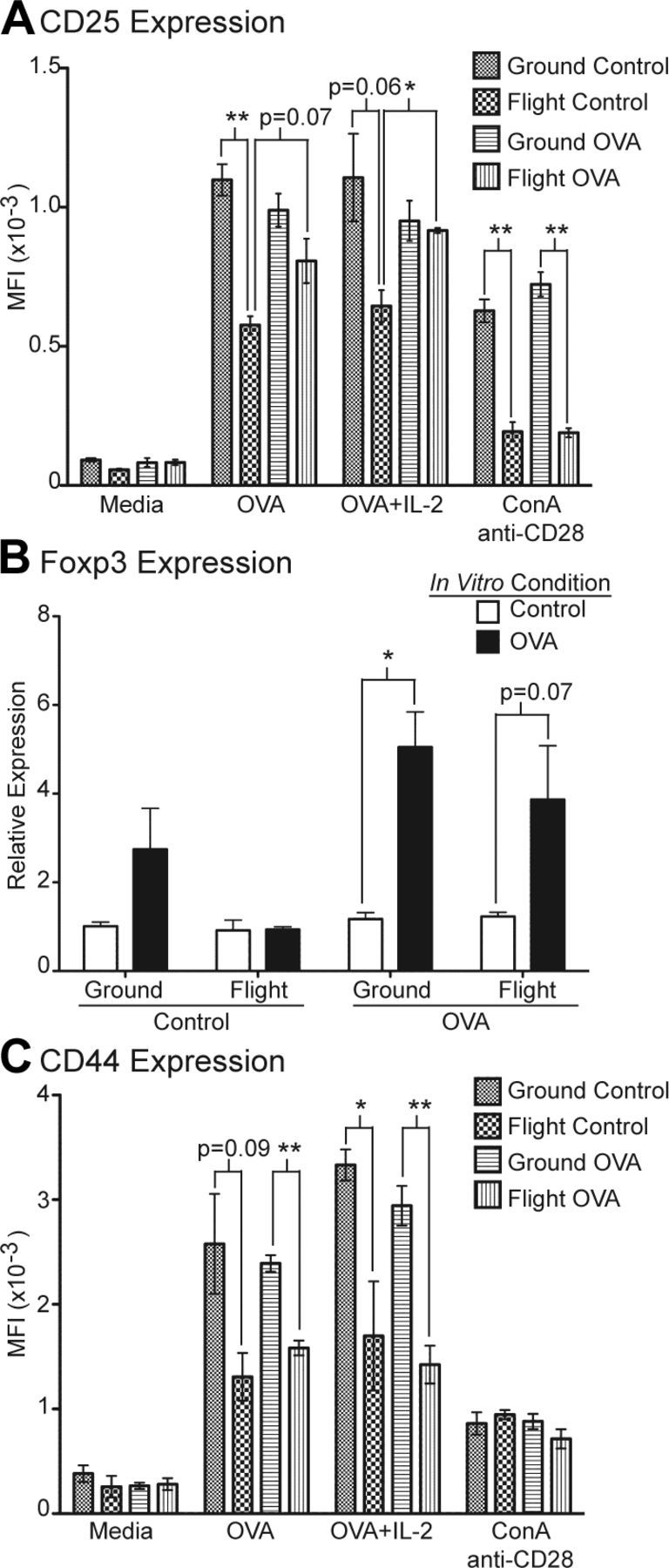
Figure 5.

Transferred OT-II cells from flight mice demonstrated impaired expression of CD25, Foxp3, and CD44 upon OVA peptide restimulation in vitro compared with ground mice. A) Splenocytes were restimulated in vitro with medium alone, OVA peptide, OVA peptide plus IL-2, or ConA and anti-CD28. After 3 d, cultured cells were stained with CD4, CD45.2, and CD25 and analyzed by FACS. Data represent MFI of CD25 for gated CD45.2+CD4+ cells for each experimental group. *P < 0.005 and **P < 0.001. B) Quantitative real-time reverse transcription-PCR analysis of Foxp3 expression in splenocytes restimulated with OVA peptide for 3 d compared with freshly collected splenocytes as control. *P < 0.005. C) Analysis of CD44 expression by FACS on CD4+CD45.2+ cells after in vitro restimulation. *P < 0.05 and **P < 0.005. All groups n = 4, except for flight control n = 3. Error bars represent se. P values were calculated by 2-tailed Student’s t test.
