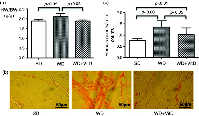
Figure 6.
(a) Heart weight/body weight ratio in the three study groups detected at the end of the study. HW: heart weight; BW: body weight. (b) Myocardial fibrosis detected by Picric acid Sirius red staining of heart sections. In the Picric acid Sirius red staining evaluation, cardiomyocytes are coloured yellow, nucleus is dark blue and collagen is fuchsia. (c) Quantification of fibrosis. The significance values are shown in the figure. Original magnification × 40; scale bar: 50 µm.

