Figure 1.
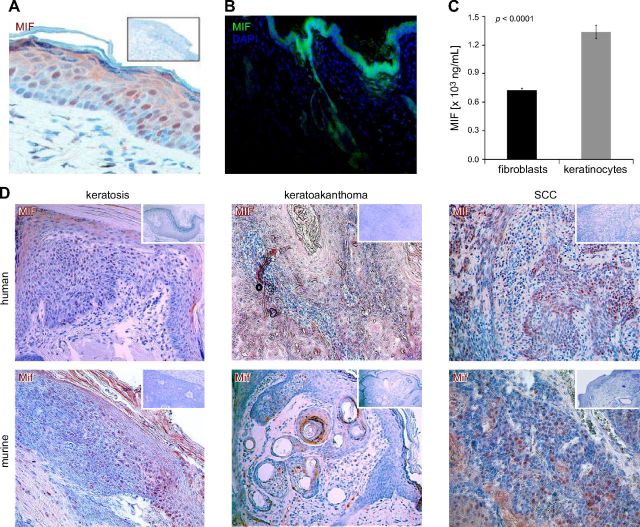
MIF expression in human and murine skin. A) MIF is constitutively expressed in untreated normal human skin. Representative section stained by IHC [3-amino-9-ethylcarbazole (AEC), reddish brown]; original magnification, ×400. Inset shows negative control staining with anti-MIF antibody omitted. Epidermal keratinocytes show more pronounced MIF staining than dermal fibroblasts. B) MIF is constitutively expressed in untreated normal murine skin. Representative section of murine skin stained by immunofluorescence. MIF expression is much stronger in epidermis and epithelial skin appendices, such as hair follicles, compared with dermal structures. MIF visualized by Alexa Fluor 488 (green), nuclei counterstained by DAPI (blue); original magnification, ×100. C) MIF secretion in growth medium by subconfluent primary human dermal fibroblasts or keratinocytes after 48 h. Keratinocytes secrete twice as much MIF as dermal fibroblasts into the supernatant. D) IHC for MIF (AEC, reddish brown) in human and murine nonmelanoma skin tumors. Top: human tumors; bottom: murine tumors; left: keratosis (precancerous lesion); middle: keratoakanthoma (benign tumor); right: squamous cell carcinomas (SCC; malignant tumor). Insets: control without anti-MIF mAb. Counterstain with hematoxylin; original magnification, ×100.

