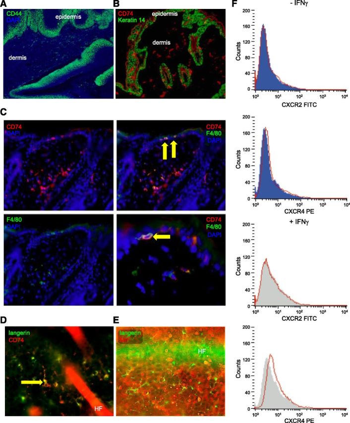
Figure 3.

Mif receptor expression and association of MIF deficiency with reduced epidermal infiltration by immune cells. A) Representative immunofluorescence for CD44 (Alexa Fluor 488, green) with DAPI nuclear counterstain (blue) demonstrating strong and constitutive expression of CD44 in the basal layers of untreated WT murine epidermis; original magnification, ×100. B) Representative IHC for expression of CD74 (Alexa Fluor 546, red) and Keratin 14 (Alexa Fluor 488, green) in skin of Mif+/+ mice. CD74 is absent on keratinocytes, but clearly expressed on immune cells infiltrating epidermis and dermis; original magnification, ×100. C) Representative double-IHC for expression of CD74 (Alexa Fluor 546, red) and F4/80 (Alexa Fluor 488, green) in skin of Mif+/+ mice. DAPI was used as nuclear counterstain (blue). Arrows show intraepidermal F4/80+ cells with coexpression of CD74. Original magnification, ×200 for all panels but lower right, which is magnified ×400. D) Immunofluorescence for langerin (Alexa Fluor 488, green) and CD74 (Alexa Fluor 546, red) with DAPI counterstain in murine epidermis demonstrating that LCs coexpress CD74. Original magnification, ×400. E) Double-immunofluorescence for langerin (Alexa Fluor 488, green) and MIF (Alexa Fluor 546, red) in untreated murine epidermis demonstrating that langerin+ cells do not express MIF. Original magnification, ×400. F) Expression of CXCR2 and CXCR4 by flow cytometry on primary normal human keratinocytes in vitro with and without prior stimulation by IFN-γ (20 ng/ml, 48 h). No expression of CXCR2 and CXCR4 is seen on keratinocytes.
