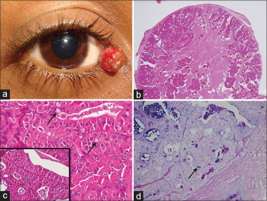
Figure 1.

Photographs from Case 1: (a) pinkish nodular mass at the inner canthus of the right eye; (b) the scanner view showing the well-circumscribed mass stretching the overlying conjunctiva and a papillary-cystic pattern (Hematoxylin and Eosin, ×20); (c) higher magnification showing three types of cells, namely, the luminal oblong oncocytes, abluminal flattened cuboidal myoepithelial, and few interspersed goblet cells (black arrow) (Hematoxylin and Eosin, ×400); the inset further highlighting the dual population of cells; (d) the goblet cells were highlighted by the blue color (black arrow) (periodic acid Schiff–alcian blue stain, ×100)
