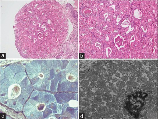
Figure 2.

Photographs from Case 2: (a and b) the mass showing a tubular/acinar pattern with eosinophilic secretion and scattered goblet cells (Hematoxylin and Eosin, ×40 and ×200); (c) the oncocytes stain blue with phospho-tungstic acid hematoxylin stain (PTAH) (×1000); (d) electron microscopy of the tumor cells showed preponderance of mitochondria with a relative diminution of other cytoplasmic organelles, some of the mitochondria having large crista (uranyl acetate and lead citrate; ×6000)
