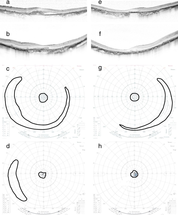
Fig. 1.
Optical coherence tomography and Goldmann perimetry data before and after 8 years of anti-VEGF therapy. Horizontal B-scan images of the left eye (a, b) and right eye (e, f) immediately before (a, e) and 8 years after (b, f) anti-VEGF therapy, respectively. Subfoveal choroidal neovascularization with serous retinal detachment was present at baseline (a). Exudative changes were well controlled and the fibrovascular membrane remained after 8 years of anti-VEGF therapy (b). Goldmann perimetry results for the left eye (c, d) and right eye (g, h) before (c, g) and 8 years after (d, h) anti-VEGF therapy, respectively. The bold lines represent V-4 isopters. The peripheral visual field was present before treatment in both eyes (d, h). However, after treatment, the peripheral visual field remained only in the left eye (c). VEGF, vascular endothelial growth factor

